Ultrasound of the Week #019
Case:
A 63 year old female presented with acute onset central abdominal pain. It had no radiation nor other red flag features and she was otherwise well, aside from AF for which she was on Warfarin.
A bedside ultrasound was performed to assess for AAA. No AAA was seen, but tenderness and some ultrasound abnormalities were noted on the anterior abdomen over her rectus sheath.
Closer examination was performed with the high frequency (linear) probe – see images below.
Question: What is identified here?
[expand title=”Answer:” tag=”h2″]
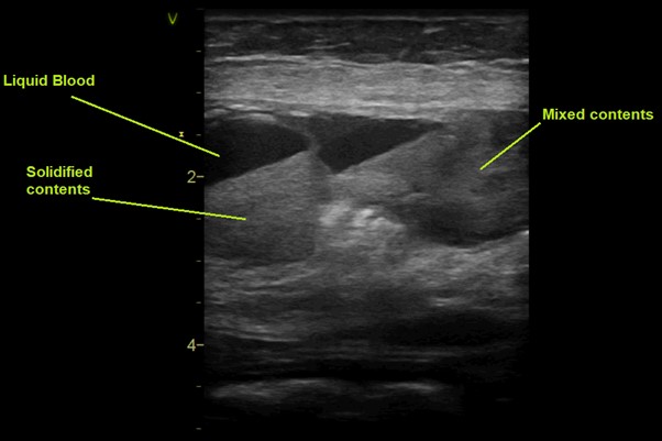
This is a longitudinal view of the rectus sheath. You can see various foci of haemorrhage at different stages. These range from liquid contents (acute – hypoechoic (black)) to more solidified haematoma (more echoic – visible in the below image with a debris/fluid level).
Haematomas often contain mixed contents of blood at various stages of coagulation, appearing as a heterogenous ill-defined masses (visible on the right of the image).
Most haematomas can be managed expectantly along with reversal of any coagulatopathy. Unstable patients or those with signs of persistent bleeding may require surgical exploration for ligation or cauterisation of bleeding vessels. This patient had her warfarin reversed in ED and was admitted under the surgical team for expectant management of her rectus sheath haematoma.