Ultrasound of the Week #018
2 Ocular cases this week, with many thanks to Dr Ahmed Abdul-Ghani for these interesting cases and explanation.
Case #1:
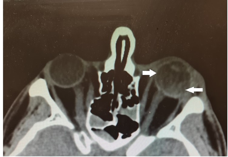
Young female presenting with migraine and a few weeks of right eye dark floaters. Visual acuity intact. No pain reported.
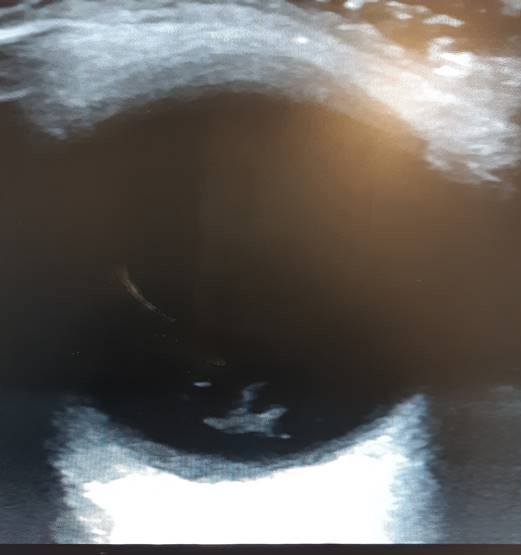
This was her ocular ultrasound – what does it show?
[expand title=”Answer:” tag=”h2″]
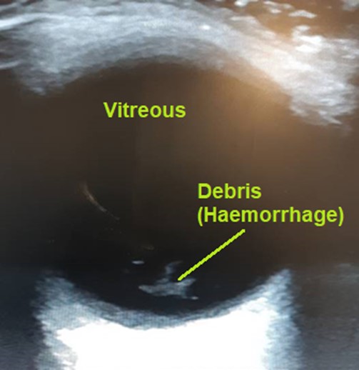
On ocular ultrasound there is echogenic debris in the posterior chamber, freely mobile on eye movements and not tethered to the RCS (Retinal-Choroid-Sclera) Complex. This is consistent with vitreous haemorrhage. In this case it was an atraumatic process with no associated retinal or posterior vitreous detachment. Ophthalmology advised conservative management.
Vitreous haemorrhage:
Vitreous haemorrhage is extravasation or leaking of blood into the vitreous humour and can result in visual floaters. The most common cause is diabetic retinopathy where extravasation of blood occurs from abnormal blood vessels.
Case #2:
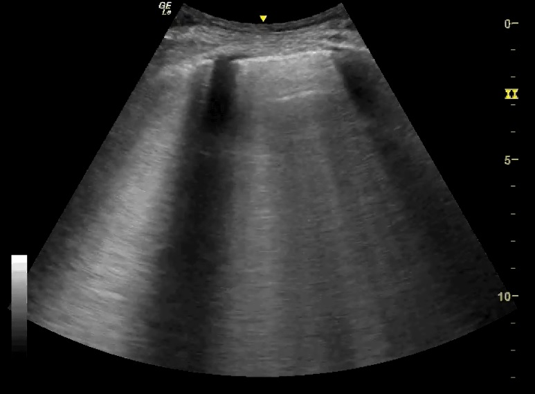
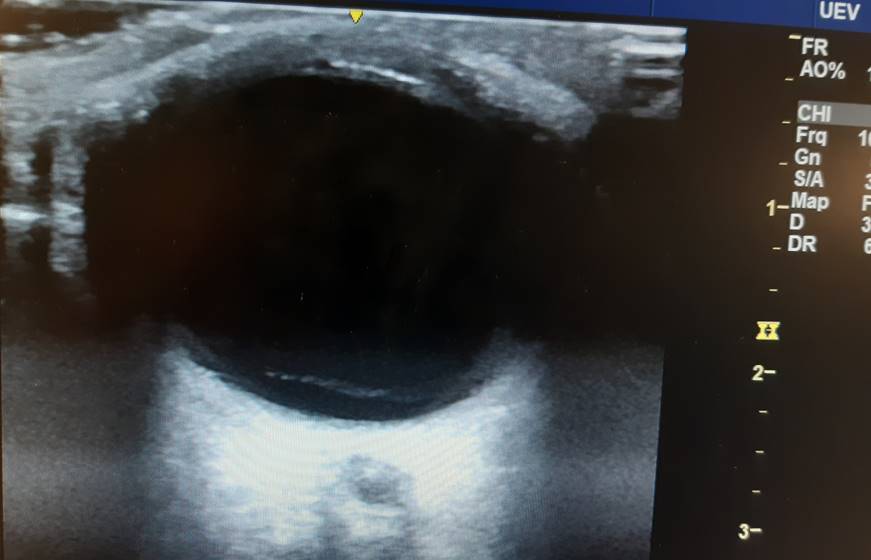
A 59 year old female presented with 10 days of painless right eye floaters and 1 day of blurred vision. She had had no trauma.
This was her ocular ultrasound – what does it show?
[expand title=”Answer:” tag=”h2″]
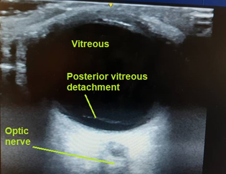
This shows a detachment flap that extends over the optic nerve. The finding is consistent with posterior vitreous detachment (PVD).
Retinal detachment (RD) can be distinguished from PVD because the RD flap will not cross past the optic nerve or ora serrata where it is anatomically tethered, whereas PVD can (see below).
Ophthalmology confirmed the diagnosis in this case – for conservative management and follow-up.
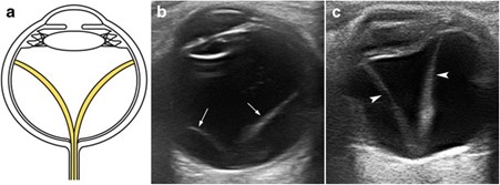
Retinal vs Vitreous detachment:
These are some examples showing Retinal detachment. Here you can see the detachment is tethered at the optic nerve. When assessing retinal detachment, a real bonus for the opthalmologists will be assessing whether the macular (lateral to the optic nerve) has become detached (‘mac on’ vs ‘mac off’), as this will affect urgency of treatment. There is a good video demonstrating this here : https://www.youtube.com/watch?v=JijIfSzOG9U
For some excellent further reading on ocular ultrasound this article has a good breakdown : https://www.ncbi.nlm.nih.gov/pmc/articles/PMC4877345/
[/expand]
References/Resources: