Ultrasound of the Week #021
Case:
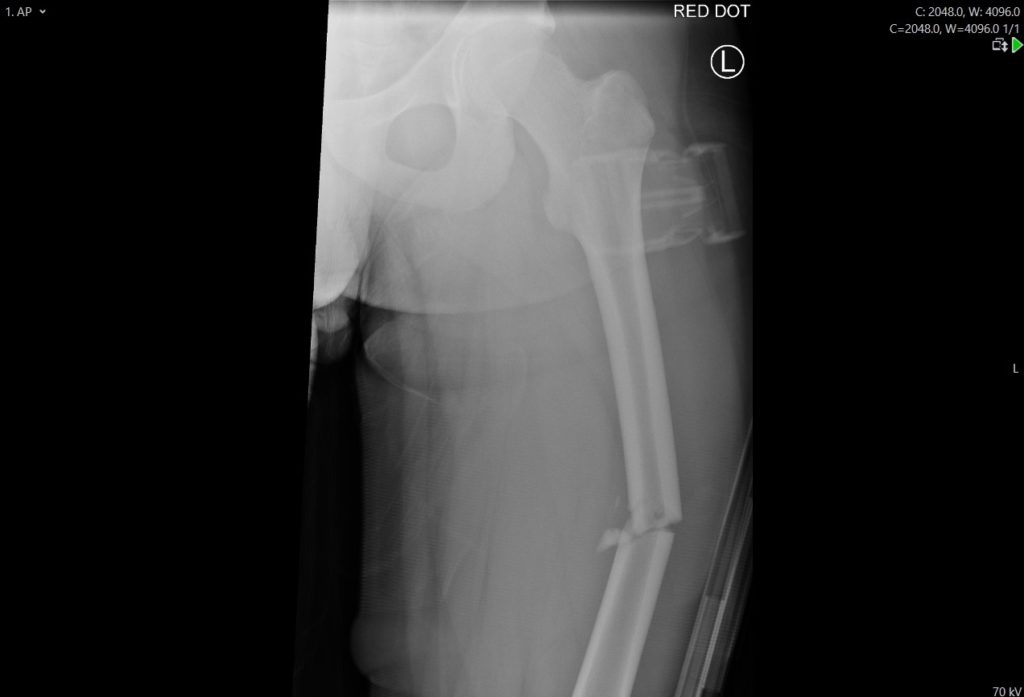
A 33 year old male who fell from 2 storeys was brought in as Code Red by HEMS. He was fixing some bird net on his balcony and fell down. On primary survey there was some left sided bony crepitus, reduced breath sounds on the left chest and left femoral and humeral fractures.
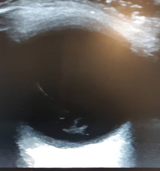
An eFAST was done at the bedside – what does this show?
[expand title=”Answer:” tag=”h2″]
There is no visible pleural sliding. This is highly suggestive of pneumothorax (although can also be seen if there is no lung movement e.g.endobronchial intubation, cardiac arrest or severe ARDS)
[/expand]This patient had a CT including facial bones and an ultrasound of the left eye.
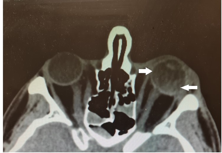
Question: What abnormality is seen here that was initially missed on CT?
In the CT image you can see hyperdense fluid in the eye (as pointed by the arrows) and in the POCUS image you can see the flaps of tissue converging on the optic disc. Another hyper resonant membrane anterior to the retina crossing the optic disc is the Vitreous membrane. This is consistent with a vitreous detachment, retinal detachment and vitreous haemorrhage and this patient was referred to Ophthalmology. [/expand]

Retinal detachment, vitreous detachment and vitreous haemorrhage are explained further in US of the Week #018. Here is a quick reminder:
Retinal Detachment:
An RD is confirmed by the presence of a bright, echogenic membrane tethered to the optic disc but separated from the choroid.
Posterior Vitreous Detachment:
A posterior VD is defined by the presence of a detached, thin, mobile membrane at the interface between the vitreous and the retina.
These 2 abnormalities are differentiated based on the visual appearance of the membrane and whether the membrane is tethered to the optic nerve.
Vitreous Haemorrhage:
A Vitreous Haemorrhage is defined by the presence of a fluid collection of variable echogenicity in the posterior chamber. Retinal detachment can be an emergency if it is ‘ Macula on’ retinal detachment and needs urgent ophthalmology intervention[3]
References:
- Alrajhi K, Woo MY, Vaillancourt C. Test characteristics of ultrasonography for the detection of pneumothorax: a systematic review and meta-analysis. Chest. 2012 Mar;141(3):703-708. doi: 10.1378/chest.11-0131. Epub 2011 Aug 25. PMID: 21868468.
- Netherton S, Milenkovic V, Taylor M, Davis PJ. Diagnostic accuracy of eFAST in the trauma patient: a systematic review and meta-analysis. CJEM. 2019 Nov;21(6):727-738. doi: 10.1017/cem.2019.381. PMID: 31317856.
- Tewari A, Shah G, Management of Primary Retinal Detachments, reviewofophthalmology.com, last accessed 10/03/2021