Ultrasound of the Week #032

Case:
A 9 year old attended with fever and vomiting on a Friday evening pre-bank holiday weekend. She had a PMH of bilateral ureteric narrowing previously managed with a right sided JJ-stent (removed 18 months ago). She had no dysuria/urinary frequency nor renal angle tenderness.
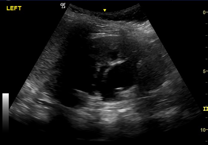
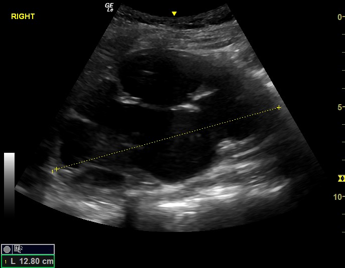
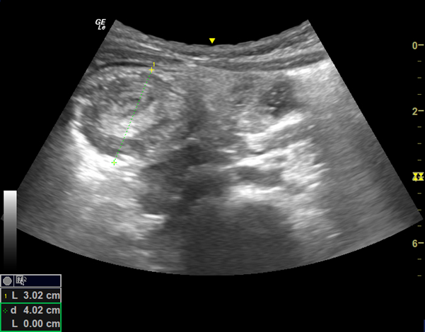
A bedside renal US showed the below.
Question: What does this show?
This shows bilateral hydronephroses, worse on the right which is severe. Comparison of her previous US KUB from 12 months earlier showed a very significant increase in the size of hydronephrosis, especially on the right side. The right kidney is larger than it should be for this age (and was indeed ballotable on examination).
This patient was admitted under the paediatric team and treated with IV antibiotics to await a formal US and urological input for a right nephrostomy.
POCUS for Hydronephrosis:
Renal US was covered previously in Ultrasound of the Week 010. Below is a short description of US for hydronephrosis and a more comprehensive review can be found here at POCUS101.
Being familiar with eFAST POCUS will provide a foundation upon which to progress to renal US. Assessment is of the degree of renal pelvis and calyceal dilatation and eventually cortical thinning.
- “Mild” hydronephrosis occurs with pelvic dilatation without calyceal dilatation.
- “Mild-moderate” is identified by the presence of pelvic and calyceal dilatation
- “Severe” is identified by gross dilatation of renal pelvis and calyces causing cortical thinning
Grading hydronephrosis is controversial and grading systems all show considerable inter-observer reliability[1]. In the Emergency Department, often a recognition of hydronephrosis with a general grading of ‘mild/moderate/severe’ is sufficient.
[/expand]References:


