Ultrasound of the Week #045
A phenomenal case this week from Dr Salman Naeem, Emergency Medicine Registrar. These are some incredible images of a relatively rare condition.
Case:
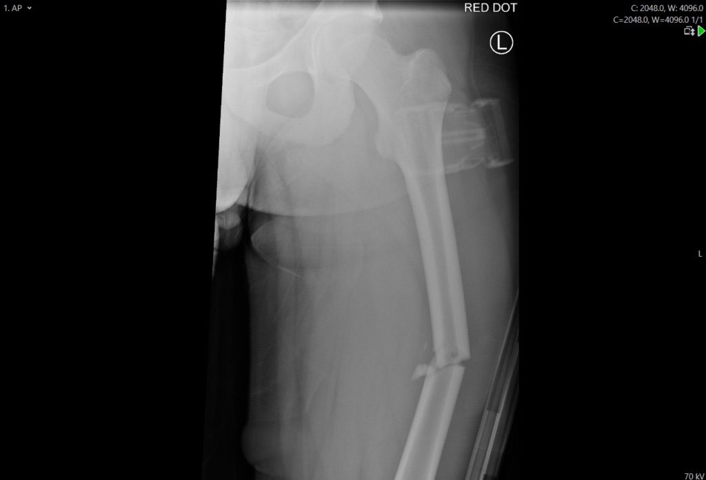
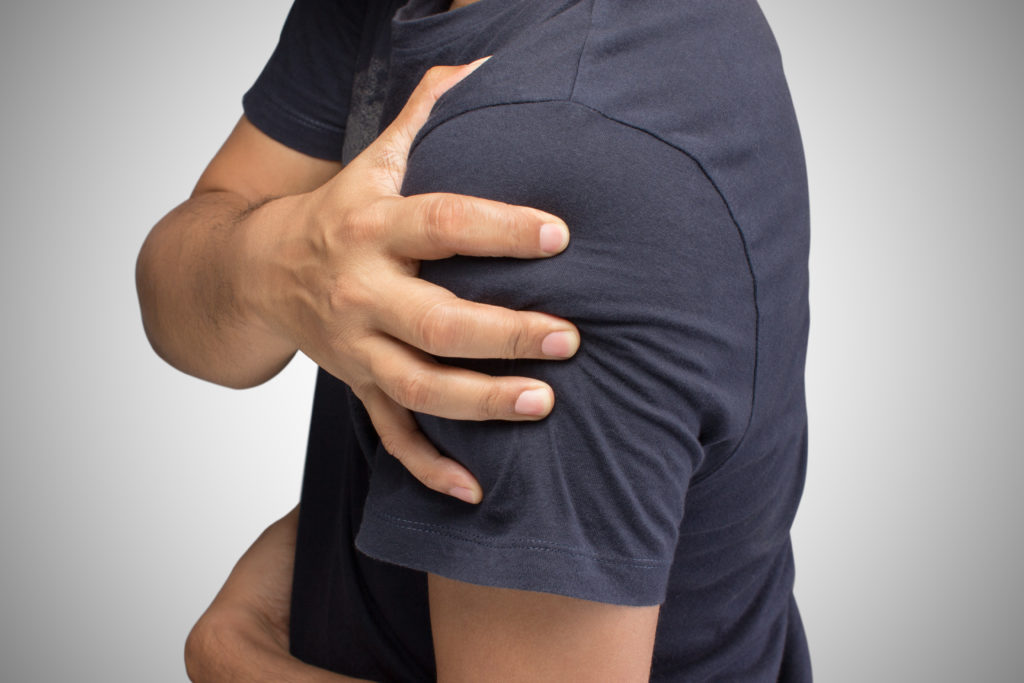
A 49 year old patient presented with worsening pain and swelling in his right shoulder and arm, two days following an assault to his face and right shoulder. He was reviewed by an Emergency Physician and the initial working diagnosis was a fracture of the right humerus and possible haemorrhagic joint effusion. He had a shoulder X-ray showing no signs of fracture. Point of Care Ultrasound (PoCUS) was performed to assess for a joint effusion and showed the following:
Findings:
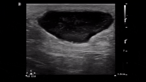
This shows a hypoechoic collection below the deltoid muscle with a 7.7mm round pulsating collection. This demonstrates the ‘ying-yang sign’ on assessment with colour doppler and can be seen to be arising from a vessel – this is the thoracoacromial artery, a branch of the axillary artery. This is a traumatic pseudoaneurysm and the pseudoaneurysm ‘neck’ can be seen on the right of the image.
Case Progression:
This significantly altered the management of the patient in prompting a CT angiogram that confirmed a leaking pseudoaneurysm in the right shoulder with surrounding hematoma. However the originating vessel could not be visualised on CTA, and the patient was admitted for an ultrasound guided thrombin injection which was successful.
Pseudoaneurysms on Ultrasound:
Traumatic pseudoaneurysms of branches of the thoracoacromial artery are very rare. Most cases reported in literature are post-infectious in aetiology. Blunt trauma can lead to weakening of the arterial wall resulting in formation of a pseudoaneurysm. Pseudoansurysms’ risk of rupture is greater than that of true aneurysms and active treatment is usually required.
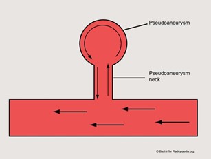
Ultrasound has been used to evaluate soft tissue swellings and pseudoaneurysms in post-traumatic injuries and intravenous drug users, and is the imaging modality of choice in psuedoaneurysms[1]. Pseudoaneurysms appear as round pulsating swellings arising from a vessel. Colour doppler shows the pathognomic ‘ying-yang’ or ‘pepsi’ sign which is due to turbulent flow of blood within the pseudoaneurysm. Remember the mnemonic ‘BART’ for direction of flow with colour doppler – Blue Away, Red Towards. Blue indicates flow away from the US probe, Red indicating flow towards the US probe.
Conclusion:
This case highlights the utility of PoCUS in the evaluation of traumatic soft tissue swellings and its ability to direct diagnostics and expedite appropriate patient management.
References: