Ultrasound of the Week #042
Case:
A tall 14 year old male presented with a few days of chest pain and breathlessness on a background of mild asthma.
[expand title=”Findings:” tag=”h2″]
Lung US: Remember – normal lung is full of air and therefore does not conduct US waves well. So if you are seeing something in the chest, that is abnormal. R1 shows dense tissue with a small rim of hypoechoic (black) matter abutting the pleura. R6 shows more hypoechoic fluid at the lung base, with dense tissue within it. The left lung image shows clear pleural sliding.
PLAX: There is a mass visible anterior to the aorta. Posteriorly to the aorta, the right pulmonary artery can be seen. This is not usually seen on PLAX and is likely only visible due to surrounding increased soft tissue which conducts ultrasound well.
Conclusion:
- No left sided pneumothorax
- Dense soft tissue in right hemithorax and mediastinum with no pneumothorax.
[/expand]
[expand title=”Case Progression & Summary:” tag=”h2″]
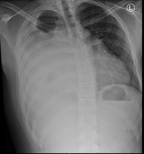
He had a chest XR performed and bloods were taken, showing a mild anaemia, WBC 13 (Neut 10) and a CRP of 144. Following this he had a departmental lung ultrasound which was reported as follows:
He was transferred to the local oncology centre for further workup and management of a likely malignant process.
Summary & Learning Points:
- POCUS is extremely useful for investigating pathology that abuts the pleura, is rapidly available and outperforms chest XR in evaluating the majority of intrathoracic pathology
- Although in this case Lung US was performed pre-CXR, any whiteout on chest XR should be imaged further with either lung US or CT.
For other Lung Ultrasound cases, see Ultrasound of the Week Lung Cases.
[/expand]