Ultrasound of the Week #029
Special thanks to Dr Dom Craver, PEM SpR for this great case and images.
Case:
A 4 year old girl came in having stepped on some broken glass at home from a broken fish tank. She had some pain in the bottom of her foot and XR showed a possible radio-opaque speck adjacent to one of her metatarsals.
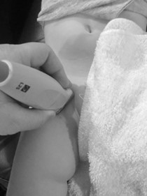
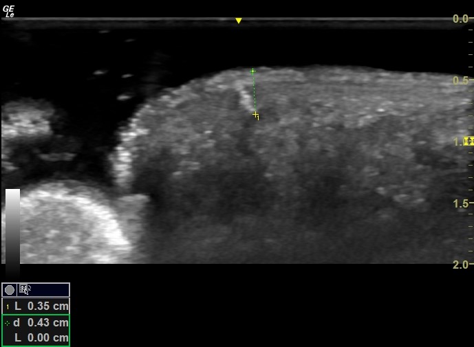
Ultrasound initially couldn’t locate a FB, but after putting her foot in a ‘waterbath’, the following image was identified.
Question : What does this show?
Answer: this shows an echogenic FB in the sole of her foot at a depth of 0.35cm.
Case progression:
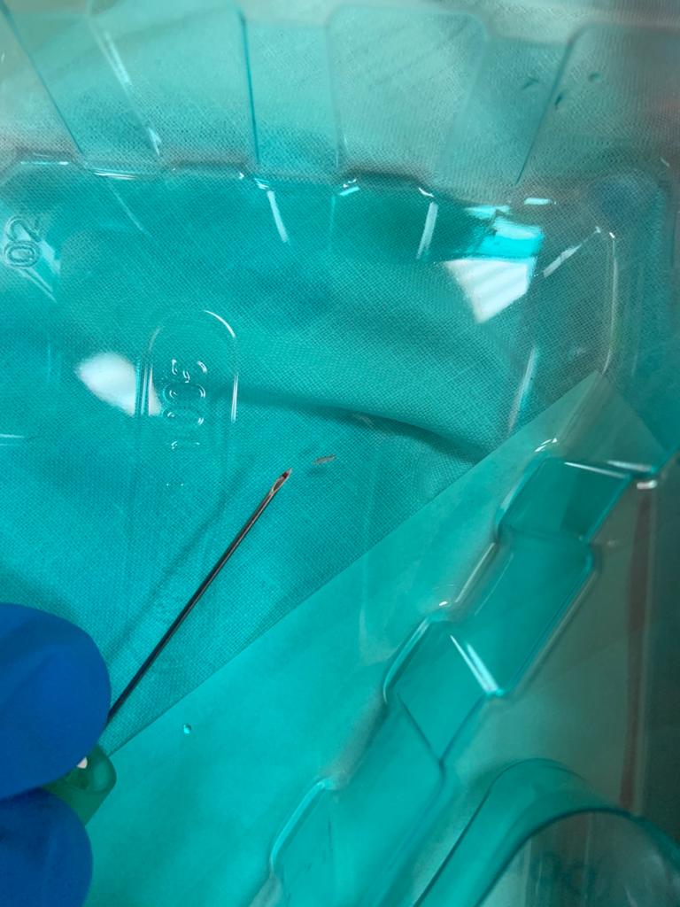
After some LMX gel and lidocaine infiltrated with an insulin needle, the following FB was removed from her foot with a needle and some splinter forceps (seen below).
POCUS for Foreign Bodies:
Foreign bodies (FB) are often difficult to locate on X-ray and radiolucent FB’s such as wood, plastic or organic material will not be seen on X-ray. However, the majority of foreign bodies are echogenic and can be visualised using ultrasound. The use of a waterbath or stand-off pad (or makeshift one using a water-filled glove or IV fluid bag) makes visualisation of superficial FB’s much easier. In this case a medical vomit bowl filled with water was used, into which the patient’s foot and ultrasound probe could be placed.
Missed FB’s can lead to ongoing pain, local infection and complaints/medicolegal claims and so POCUS for FB’s is an excellent tool for use in the ED. There is a great review with many examples available at https://www.acep.org/sonoguide2/procedures/foreign-bodies/.
References/Resources: