Ultrasound of the Week #028

Thanks to EM SCF Dr Thomas Durrand and US Fellow Dr Salman Naeem for this great case.
Case:
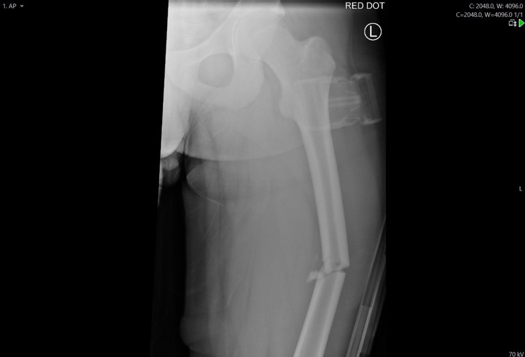
A 97 year old male was brought into resus as a trauma call, having fallen down stairs at home. He complained of left sided chest & pelvic pain. He had a known PMH of myelodysplastic syndrome, previous EVAR and AF yet was not anticoagulated.
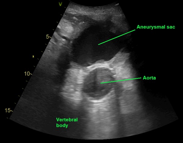
He was taken for a trauma CT and a POCUS scan of his abdominal swelling was performed.
Question : What does this show?
[expand title=”Answer:” tag=”h2″]
This is a leaking Abdominal Aortic Aneurysm (AAA), identified as a pulsatile structure to the left of and anterior to the vertebral bodies. In this case the previous EVAR graft has ruptured and blood is leaking from the true lumen into the aneurysmal sac, demonstrated beautifully here on colour doppler.

This allowed for rapid discussion with Vascular and Interventional Radiology teams, illustrating the extent of the bleed and facilitating discussions regarding management options. After some discussion, it was agreed with the team and patient that palliative management would be best and it was arranged for him to be transported home.
POCUS for AAA (RCEM Core Competency):
POCUS is an excellent modality to assess for AAA. It is quick and easy to perform and is taught as one of the Core RCEM Level 1 modalities. The aorta is scanned from the coeliac axis down to the bifurcation into the common iliac arteries. Three Anterioposterior (AP) measurements should be made and in the acute setting an AAA is defined as a transverse diameter >3cm[1]. If the aorta is visualised throughout and is <3cm, an AAA can be excluded, making it an excellent ‘rule out’ test, with a NPV of 98.6% and PPV of 100%[1].
For further reading and educational resources on AAA scanning and other Level 1 modalities, there are some great resources on the RCEMLearning website at https://www.rcemlearning.co.uk/curriculum-ultrasound/.
[/expand]
References/Resources:


