Ultrasound of the Week #001
Case :
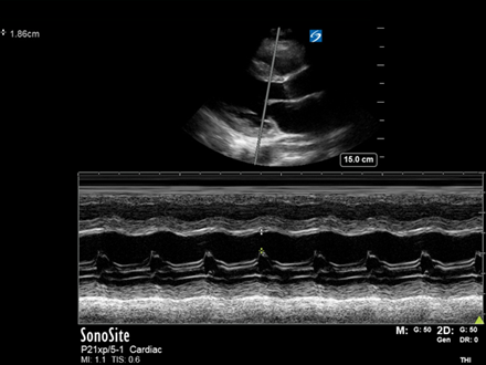
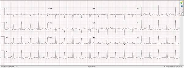
A male in his 30’s seen by LAS with chest pain and ST elevation on his ECG was taken to the local PCI centre. Here he had a rapid echo done showing no Regional Wall Motion Abnormalities (RWMAs) suggestive of acute MI. The cardiology team then transferred him to their corresponding Emergency Department for further assessment.
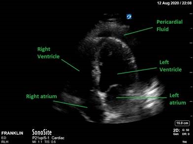
The EM Registrar saw the patient and performed a focused bedside echocardiogram showing the below image:
Question : What is the obvious abnormality? How might this account for the patient’s symptoms?
Answer
Case Progression :
The patient had signs and symptoms consistent with pericarditis and was treated as such and referred to the medical team for monitoring. This effusion & patient didn’t show any signs of pericardial tamponade – we will cover images and echo findings of tamponade in a future case.
This demonstrates that even specialists miss things – always have a look at your on X-ray’s, CT’s and ultrasounds!