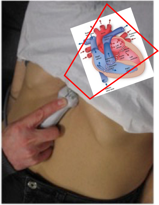
Subxiphoid View (SXV):
The SXV is an excellent view for a quick assessment of general heart function and particularly good for pericardial fluid. It is generally the easiest echo window to learn and perform quickly.
Obtaining the View
Use the cardiac OR abdominal probe with the probe marker to the patient’s right. Set the starting depth to 15 – 20cm. Position the probe in the epigastrium, just caudal to the xiphisternum & ribs, aiming under the ribs and slightly towards the left shoulder. Press down gently in a flattish plane to aim up and under the xiphisternum.
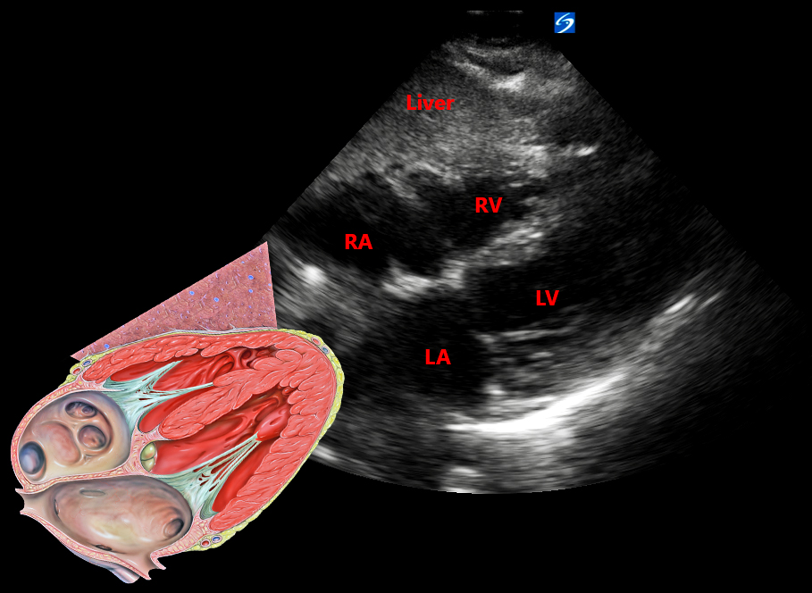
Although the probe is positioned below the heart looking up, the view obtained is inverted as a triangular shape projecting from the top of the tablet screen expanding outwards and downwards. The top of the screen represents the tip of the ultrasound probe. The view will resemble the image below with the heart chambers as labelled. The RV and RA are ‘shallow’ as they are close to the probe, with LV/ LA ‘deeper’ in the image (further away from the probe). The liver may be appreciated in front of the RV. Centralise the image and adjust the size and gain to see the whole heart (usually just unable to accommodate the apex). Any pericardial fluid should be easily seen in this view.
An SXV view of the heart should be achieved and then reviewed from top – bottom evaluating the following:
- Is there a pericardial collection? (May be noted superiorly between the liver and the RA/ RV initially, as it increases in quantity, circumferentially around the heart, or inferiorly around the LA/ LV. Fluid on ultrasound is black (anechoic). There may be compromise of RA/ RV movement).
- Look at the Right Ventricle
- Is the right ventricle normal size or dilated?
- Is the right ventricle contracting well or poorly?
- Look at the Left Ventricle
- Is the left ventricle empty or full?
- Is the left ventricle contracting well or poorly?
Troubleshooting
The SXV may be difficult in those with distended or tender abdomens and in patients that are sitting up or have high BMI.
- Start with reasonable depth (±15 – 20cm)
- Move a little to the patient’s right to use the liver as a ‘window’ as it conducts ultrasound well.
- Ask the patient to breathe in slowly and hold their breath briefly where you get the best image. (This pushes the diaphragm and heart lower and closer to the ultrasound probe)
- Flex the knees of the patient. This allows the rectus muscles to relax and facilitates pushing the probe up under the xiphisternum.
Video Review: