Two specific examinations are valuable to identify haemorrhage. A bilateral evaluation in the most basal (dependent) areas of the chest (depending on patient position) evaluating for presence or absence of a large fluid collection; and the right upper quadrant (RUQ) view of a Trauma FAST Scan evaluating for a fluid collection in Morrison’s pouch or around the liver; in order to answer the following questions:
- Is there the presence of a massive haemothorax?
- Is there the presence of a massive haemoperitoneum?
Haemothorax:
Ultrasound has been shown to be sensitive enough to detect ± 10 – 20ml of pleural fluid. In the context of trauma this represents haemothorax until proven otherwise. Clinically significant haemothoraces in the pre-hospital setting should be easily visualised.
Obtaining the View
Place the abdominal probe in the mid-axillary line at the level of the costal margin with the marker towards the head. Set the depth to 10 – 15cm.
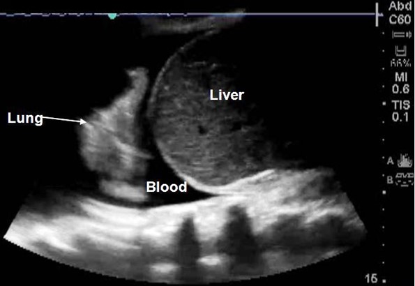
Anatomy to identify is the liver and diaphragm (bright white line). Look above the diaphragm and aim to identify lung and a large dark collection of fluid.
Fluid in the pleural space appears anechoic (black) and above the diaphragm. Collapsed lung may be seen floating in fluid.
Haemoperitoneum:
Blood is most often found on a Trauma FAST Scan in the RUQ area in patients with multiple intraperitoneal injuries or isolated injury to the liver, spleen, or retroperitoneum. Of the three areas visible in this view (hepato-diaphragmatic, Morrison’s pouch, caudal liver edge) the caudal liver edge is the area most sensitive for the detection of blood.

Obtaining the View
Keep the abdominal probe in the anterior to mid-axillary line and move inferiorly towards the feet using the diaphragm as a landmark. Note any black(anechoic) shadows between the diaphragm and liver (hepato-diaphragmatic), between the liver and the kidney (Morrison’s Pouch), and the lower edge of the liver
Move the probe caudally towards the feet aiming to visualise the whole kidney including the lower pole.
The absence of free fluid on US does not rule out peritoneal haemorrhage and serial scans should be considered in high risk patients.
Video Review: